Health and Life Sciences
How neutron scattering can contribute to life sciences and health
The increase of life expectancy, the growth of the world´s population and the changes in the Earth´s climate mean that we are today faced with major health challenges. In recent years there have been spectacular advances in the understanding of the molecular and cellular basis of disease, thanks to fundamental discoveries in chemistry, physics and biology. Fighting disease doesn't only involve developing treatment, but also relies on a better understanding of fields such as genetics, environmental factors and nutrition.
Research in diverse areas is being carried out today that may have an impact on the future of the world´s population health: not only biomedicine but also food science, water science, environmental science, materials science and biotechnology. Molecular-level information is crucial in all of these areas. It is becoming increasingly obvious that the most effective approaches involve the combined use of a wide range of techniques linking length scales from the atomic/molecular levels to macromolecular levels at which function/properties are manifested. Neutron scattering is an invaluable tool that can make vital contributions through the provision of novel information that cannot be acquired using any other technique.
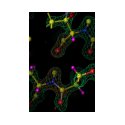
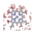
Neutrons can see the elements and molecules of life in a way that is not possible using X-rays – for example critical detail is provided on hydrogen bonding and hydration, which are aspects of macromolecular systems that are critical to biological function and enzymatic action. This type of information on biological structures and on the pathways associated with protein assembly (or mis-assembly) is critical to understand the molecular and cellular basis of disease. For example, neutron scattering data are providing important structural information of relevance to degenerative diseases such as Alzheimer's disease and other conditions that are associated with the deposition of insoluble fibrils in the body.

Neutrons shed light on how to make pregnancy tests more sensitive and cheaper. Click on the image to learn more!
Neutron techniques are also being used to study drug molecules and their interactions with a variety of biological molecules. Such studies are of interest in drug discovery and delivery and may result in new therapeutic approaches in the future. Neutrons are also used for the production of radionuclides that are used in medical diagnosis. There is also increasing interest in the development of approaches whereby neutron beams can be used to target and destroy tumours.
Neutrons are gentle probes and penetrate materials easily without damaging them. They are very precise and extremely sensitive to the detailed structure of synthetic materials. They are powerful tools for the characterisation of new materials – for example recent neutron scattering work is providing data that are of central interest for the development of new biomaterials for dental and bone implants.
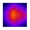
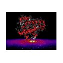
Key neutron techniques in this area include Neutron Small Angle Scattering (SANS), which provides information on the dimensions, shapes and morphology of large molecules and macromolecular complexes. Neutron Macromolecular Crystallography (NMX) gives unique structural information at high resolution, inclusive of vital detail relating to hydration and protonation states of key functional areas of proteins. Neutron Reflection (NR) can be used to study interfaces such as surfaces and membranes. Other areas such as inelastic neutron scattering and elastic incoherent neutron scattering can reveal important aspects of molecular motion. Neutron imaging methods are powerful tools for non-invasive in-vivo imaging.
Further reading
- Neutrons shed light on how to make pregnancy tests more sensitive and cheaper
- New humidity chamber will shed light on processes behind Alzheimer’s disease
- To broaden research possibilities: a new pressure cell
- Healthy diet? Using neutrons to quantify selenium in cereal crops, NMI3 article
- Assessing exposure to pollution in industrial workplaces
- Towards a new tumour-specific contrast agent for MRI applications
- Neutron scattering for the characterisation of magnetic fluids for anticancer treatment
- For more information about neutrons and biological research, please see here.
Health and Life Sciences picture gallery


