Tasks and partners
Neutron imaging for materials science – micro and nano structures
Contact: Nikolay Kardjilov, HZB
Task 1: Nano- and micro structures resolved dark-field neutron imaging with grating interferometers
Partners: PSI, HZB, FRM-II
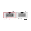
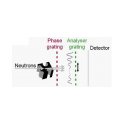
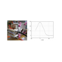
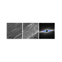
Up to now the grating interferometric technique is used for phase contrast imaging and magnetic imaging. There are few feasible studies which show the potential of the method for microstructural analysis. In this case dark-field imaging provides results which are a qualitative comparison to a position sensitive (U)SANS approaches. An example of testing of fine grained marble by dark-field radiography is shown in the figure below.
Task 2: Direct high-resolution imaging
Partners: FRM II, PSI, HZB, NPI
Recent developments of new detector systems allow for improving the spatial resolution in neutron imaging experiments down to 20 µm. Further improvement of the optical components and using of new type of neutron detection techniques like detectors based on borated micro channel plates can provide resolution below 10 µm. This will help for visualisation of structural micro details in 2D images and 3D tomographic volumes. Additionally dedicated neutron optical components like focusing neutron guides, refractive lenses and dispersive monochromators based on double reflection process will improve the beam collimation and provided the necessary intensity.
Application areas are investigation of micro cellular materials like metal and polyester foam structures (Partners: TU Berlin, University of Valladolid), porous materials like Membrane Electrode Assemblies (MEA) and GDL systems in PEM fuel cell research (Partners: ZSW, University of Ulm, FZJ) and wood research (Partners: University of Palermo, PSI, ETH-Zürich).
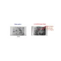
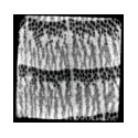
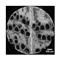
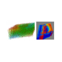
The pictures below show a micro-tomography investigation of an oak wood sample with neutrons (left) and synchrotoron radiation (right) – both measured at PSI – indicating the lack in resolution obtained with neutrons. Because neutrons are very sensitive for water, its use for non-destructive testing with much higher resolution will be one aim in the project in order to contribute to modern wood research and plant’s physiology.
Task 3: Strain and stress mapping
Partners: HZB, FRM-II, PSI, ISIS
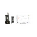
The most common method to determine stress and strain in engineering materials is neutron diffraction techniques which involve measuring atomic spacing of target planes using small gage volumes, with a present day limiting volume resolution of 0.5mm3. Energy selective imaging has the potential to significantly overcome this limitation providing a spatial resolution of 50 µm.
The method should be optimized for high spatial resolution of residual stresses in metallic samples. Areas of applications are residual stress accumulation and annealing, analysis of fatigue tests, optimization of welding techniques (e.g. Friction Steer Welding) and various industrial inspection procedures.
The energy selective method can be used very successfully for material phase separation by choosing the neutron wavelength to be between the Bragg edges of the two material phases (e.g. γ- and α – ferrite). In this case a volumetric phase separation can be achieved if tomographic experiment will be performed. This feature is very important for investigation of hardening of steels with high spatial resolution.
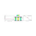
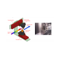
Reference for the figures below: R. Woracek, D. Penumadu, N. Kardjilov, A. Hilger, M. Strobl, R.C. Wimpory, I. Manke and J. Banhart, J. Appl. Phys. 109, 093506 (2011); http://dx.doi.org/10.1063/1.3582138.
Imaging of magnetic micro and nano-structures
Contact: Frédéric OTT, LLB
We propose that neutron scattering techniques can be extended to perform vectorial magnetic imaging in the volume of nano and micro-structures. We propose to explore 3 complementary routes which will be aimed at 3 different types of systems:
Task 4: SANS 3D: vectorial magnetic imaging of nano-particles
Partners: CEA, Jülich, STFC, TUM, TUD
There is an obvious interest for the determination of the magnetic configurations in these particles. However, the existing magnetic imaging techniques (X-ray PEEM, AFM, electron holography) are mostly surface techniques and they do not provide direct information about the volume magnetization of nano-objects.
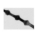
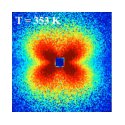
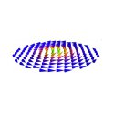
In this task we propose to develop the tools to exploit the technique of SANS with polarization analysis in order to study the magnetization distribution in magnetic particles of complex geometrical shapes.
During the last few years polarized SANS with polarization analysis has been made available thanks to the use of 3He polarizing cells. The technique is presently available at a few facilities. However, the potential of SANS with polarization analysis has until now only been scratched mostly because the technique has been made available only recently and because the tools to exploit the data do not exist.
In order to extend the accessible Q-range to small Q-values, the possibility of implementing polarization and polarization analysis in Multiple SANS will be demonstrated (FRM II – PSI). This would enable to extend the technique to micron-scale magnetic structures (as typically found in magnetic domain formations for example). This technique could bridge the length-scale gap between classical SANS and tomography.
Spherical polarization analysis is a tool which has been developed during the last 2 decades for single crystal diffraction. The use of spherical polarimetry will be evaluated for SANS experiments on small magnetic crystals in which long range magnetic modulations appear (such as in magneto-electric crystals for example). CRYOPAD will be used by TUD for such experiments.


Task 5: Precessionnal spectroscopy: vectorial magnetic imaging of planar structures.
Partners: LLB, JCNS, ISIS
Besides magnetic nanoparticles, there is an interest in probing the magnetic structure of planar systems (films) in which the magnetization is not homogeneous. As mentioned before, the existing magnetic imaging techniques such as AFM, Kerr imaging, x-ray PEEM are surface techniques and usually only probe the upper 10nm of magnetic systems which often need to be specifically designed for these studies. On the other hand, a wealth of film systems is produced by the magnetism community. Neutron reflectivity is a technique which has proved very efficient in probing these systems.
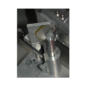
In this task, we propose to develop a new technique which could be complementary to polarized neutron reflectivity and allow probing buried non collinear structure with typical length-scales in the range 100nm-1µm.
Task 6: Direct magnetic neutron imaging
Partners: HZB, PSI, FRM2
The aim of this task is to push the limits of direct magnetic imaging in order to be able to resolve magnetic structures at the µm scale (in the direct space). This can be done by implementing advanced neutron optics as developed within the WP17 of the NMI3 consortium.
By using of standard neutron imaging detector based on CCD camera a spatial resolution of 20 µm was achieved. Further optimisation of the detector components – new scintillating screen and lens system can help to improve the resolution down to 6 µm (first experiments in cooperation with ESRF were already performed). The problem in this case is the limited neutron flux which reflects in very long exposure times. In order to solve the problem focusing neutron optics can be used for producing of high intensive neutron spot of size of few millimeters.
